CT
 CT
CT
CT
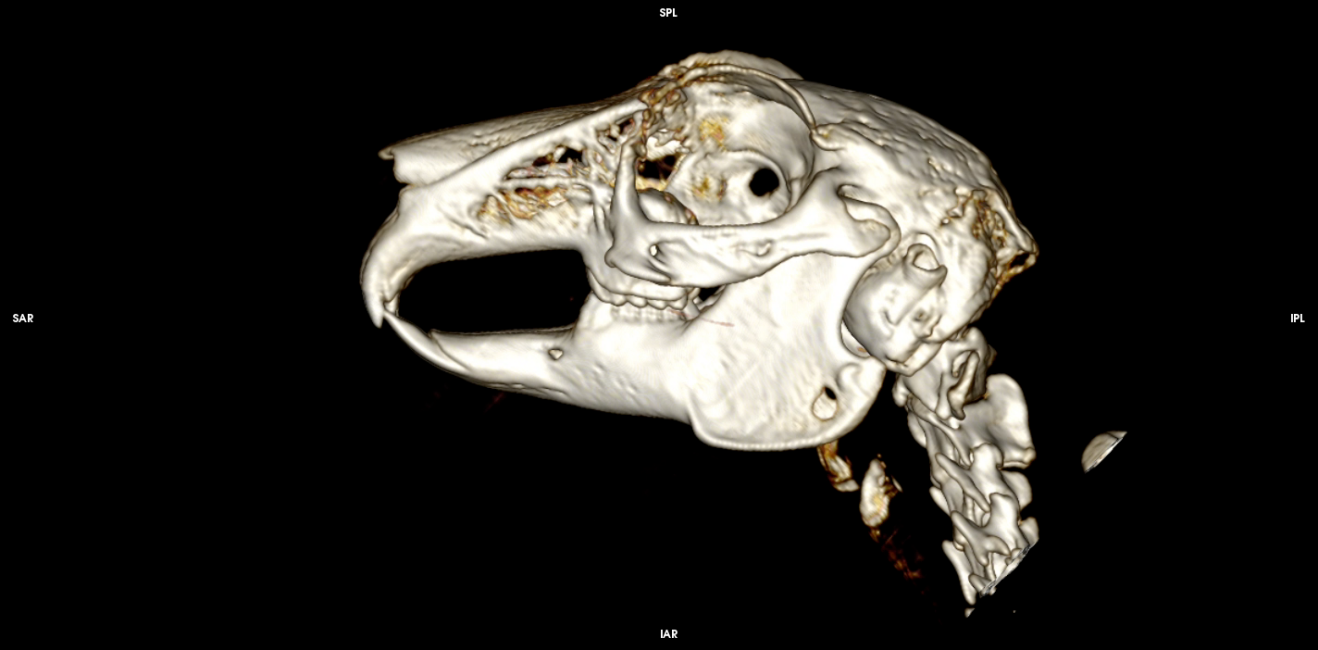
Head study
CatThin-slice CT of the cranium and sinonasal, orbital and intracranial structures.
CT
 CT
CT
CT
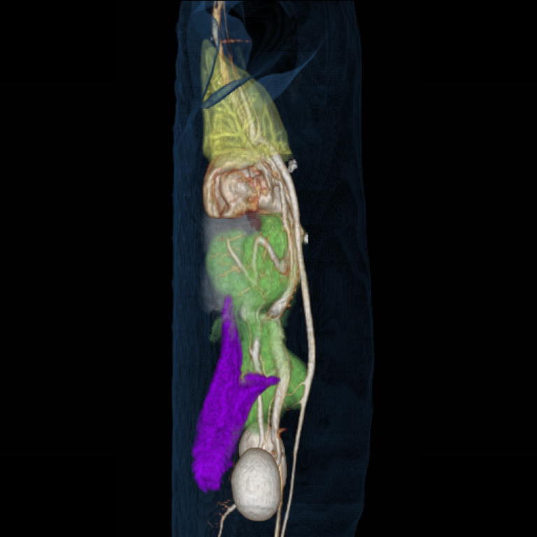
Diaphragmatic pericardial hernia (3D)
DogVolume-rendered 3D reconstruction delineating herniation of abdominal viscera into the pericardial sac.
CT
 CT
CT
CT
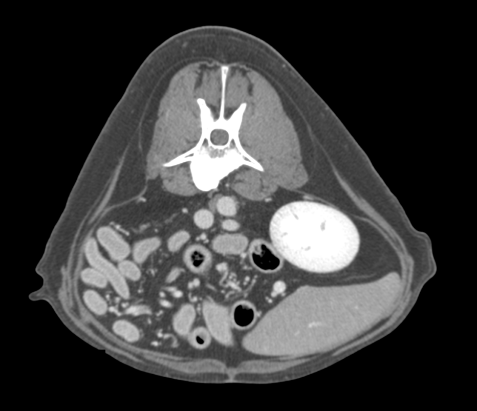
Abdominal study (contrast-enhanced)
CatContrast-enhanced cross-sectional imaging of the abdominal organs.
CT
 CT
CT
CT
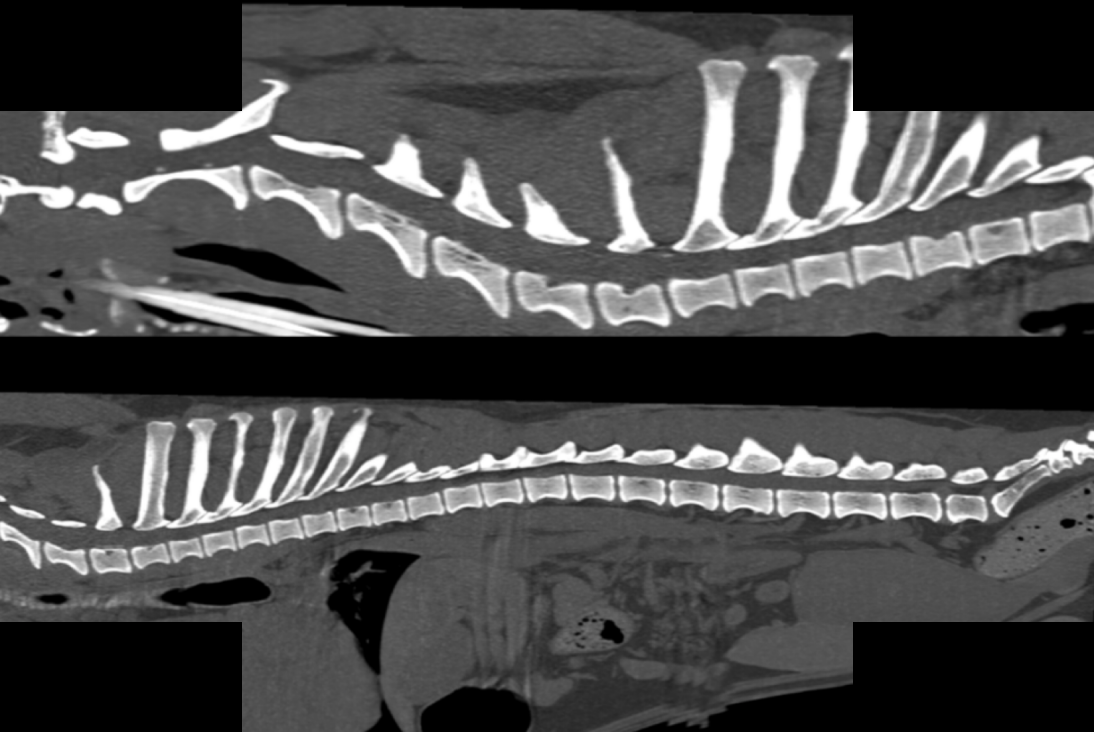
Spine study
DogHigh-resolution imaging of the vertebral column and spinal canal.
CT
 CT
CT
CT
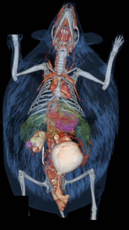
3D reconstruction
HedgehogVolume-rendered 3D reconstruction of the hedgehog’s skeletal and integumentary anatomy.
CT
 CT
CT
CT
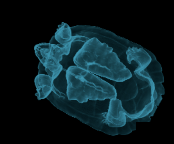
Lung study (3D reconstruction)
Turtle3D reconstruction of pulmonary anatomy within the carapace.
CT
 CT
CT
CT
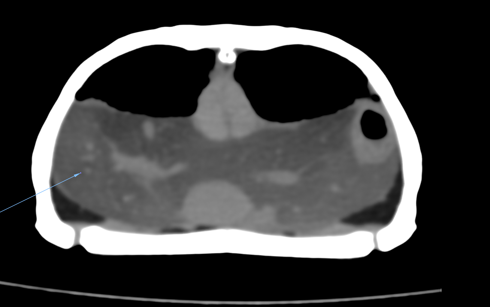
Fatty liver (hepatic lipidosis)
TurtleDiffuse low-attenuation of the hepatic parenchyma on transverse CT, consistent with hepatic lipidosis.
CT
 CT
CT
CT
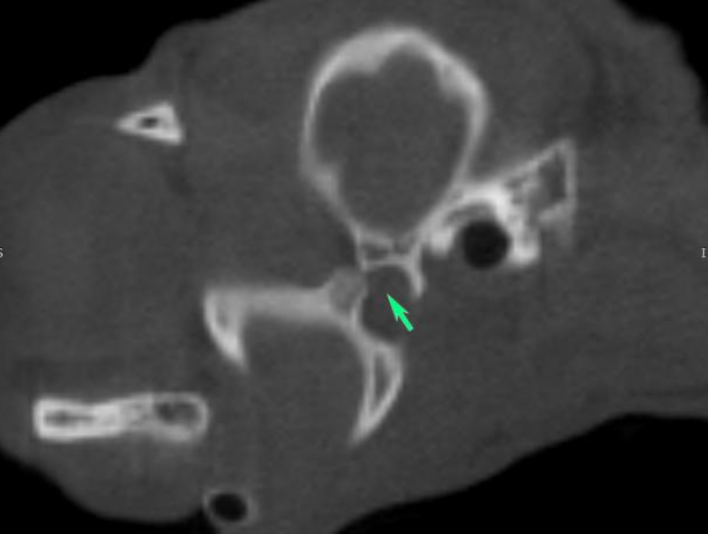
Temporomandibular joint luxation
CatTransverse CT showing rostral luxation of the mandibular condyle with an empty glenoid fossa.